Fluoroscopy
Fluoroscopy Studies includes but not limited to the following exams:
Barium studies used to evaluate the GI tract from mouth to rectum utilizing oral contrast.
- Barium Swallow: evaluates the pharynx and esophagus including the swallowing mechanism. It includes conventional barium esophagram (barium swallow) and modified barium swallow (oral and pharyngeal function study).
- Upper Gastrointestinal Series: An upper GI tract exam is a study to enable the radiologist to examine the anatomy and function of the pharynx, esophagus, stomach and the duodenum. The procedure is performed using xray in motion (fluoroscopy) after the intake of oral barium contrast. Your physician may order an upper GI exam to check for: Ulcers, tumors, inflammation, hiatal hernias, gastroesophgeal reflux disease (GERD), cause of obstruction which can lead to unexplained vomiting or bleeding etiology.
- Small Bowel Follow-Through: used to evaluate the small intestines after drinking oral contrast. The radiologist looks for cause of obstruction, inflammation, strictures, or masses.
- Barium Enema: used to examine the large intestine or colon. The test looks at the ascending colon, transverse colon, descending colon and the rectum. The radiologist may able to diagnose ulcers, benign tumors, polyps, cancer or other colonic disorders. The procedure is helpful in assessing for: Chronic diarrhea, blood in stools, constipation, unexplained weight loss and inflammatory bowel diseases, including Crohn’s disease and ulcerative colitis.
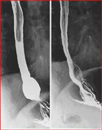
Esophagram
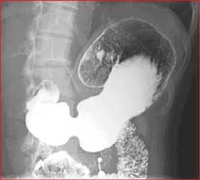
Double contrast upper GI
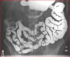
Small bowel follow-through

Double contrast barium enema
Intravenous Pyelogram (IVP):
An intravenous pyelogram (IVP) is an xray examination of the kidneys, ureters, urinary bladder and urethra that uses intravenous contrast material. When a contrast material is injected, it reaches the kidney via the blood stream. A series of X-ray pictures is then taken at timed intervals. An IVP can show the size, shape, and position and function of the urinary tract, and it can evaluate the collecting system inside the kidneys. It is commonly performed to identify diseases of the urinary tract, such as kidney stones, tumors, infection or structural abnormalities. The exam can help diagnose symptoms such as blood in the urine or pain in the side or lower back.
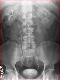
IVP
Hysterosalpingogram (HSG):
A hysterosalpingogram or HSG is an x-ray exam performed to determine whether the fallopian tubes are open and to evaluate the shape of the uterine cavity. It often is done for women who are having a hard time becoming pregnant (infertile). During a hysterosalpingogram, a contrast material is injected through a tiny tube that is placed through the vagina and into the uterine cavity. Multiple images are taken using fluoroscopy as the contrast passes through the uterus and fallopian tubes. It is usually done after menses have ended, but before ovulation, to prevent interference with an early pregnancy.
A hysterosalpingogram is usually done to assess for blocked fallopian tubes. Prior infection can cause severe scarring of the fallopian tubes leading to blocked tubes which in turns prevent pregnancy to occur. Occasionally the dye used during a hysterosalpingogram will push through and clear a blocked tube. It also evaluates the uterus for structural abnormalities such as polyps, fibroids, adhesions, or a foreign object. It is also used to assess whether surgery to reverse a tubal ligation has been successful or to evaluate the appropriate blockage of the tubes by tiny metal coils used for birth control purpose.
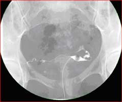
Normal HSG
