Diagnostic Oncology and PET/CT Imaging
A dual-purpose imaging device, PET/CT is literally the combination of PET (positron emission tomography) and CT (computed tomography) imaging techniques within a single machine. A PET exam detects metabolic or chemical activity in the body – how your cells are utilizing nutrients like sugar and oxygen. A CT scan uses a combination of x-rays and computers to provide the radiologist a non-invasive image of the body's anatomical structures. Therefore, PET scan would highlight a tumor's increased glucose consumption, while a CT scan would reveal its physical mass. The individual scans, which are taken virtually simultaneously, can be presented separately or as a single, overlapping, or "fused" image in order to provide the most complete information on cancer location and metastasis better than any other imaging study.
Physicians order the test to uncover a suspected cancer that has not been shown using more conventional modalities, to stage cancer, monitor treatment and determine if a treatment is working. PET/CT fusion imaging is most valuable for lung cancer and cancers located in regions of the body that have a complicated anatomy, such as the neck and lower pelvis. These locations of the body contain organs, tissue, muscles, bones and lymph nodes, all in close proximity—making the precise overlay of PET and CT remarkably helpful. PET/CT imaging has been proven to be the modality of choice in diagnosing and following up the following malignant cancers: lung, eshophagus, colorectal, lymphoma, melanoma, breast, head and neck, thyroid and cervical cancer.
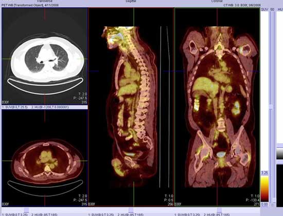
Fused PET/CT Image